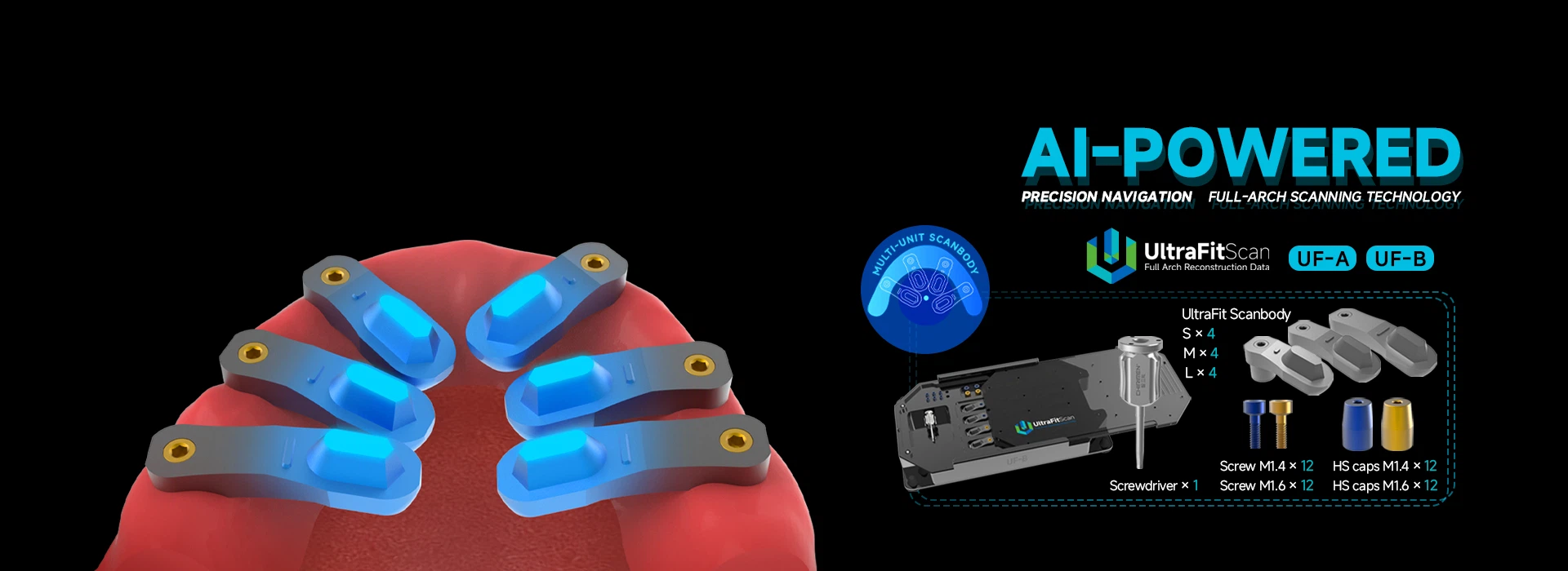


Scan Bodies For Implant
Description
Technical Parameters
Production Introduction
scan bodies for implant is the Dental Lab Scan Body Compatible with Astra Tech Ev Implant System by Abutment EV/4.2. and Chirimen can provide dental smart digital solutions.
Hiossen scan bodies for implant has HG Regular and HG Mini Blue Light Scanning Body. The Hiossen HG Regular Scanbody is part of the Scan Buddy 10 Pack, Everything you need to scan all 10 restoration options from Chirimen to the Digital Choice Library.
The Hiossen Mini Scan Body is part of the Scan Buddy 8 Pack, Issue 02. Everything you need to scan Chirimen's latest 8 restoration options through the Digital Choice Library.
Key points:
1. desktop use
2. Made in China.
3. It is manufactured by Shenzhen Chirimen Technology Co.,Ltd.
Technical specifications
Package dimensions | 2.87 x 1.97 x 0.47 inches; 0.35 Ounces |
product weight | 0.352 ounces |
Manufacturer | Shenzhen Chirimen Technology Co.,Ltd |
Product model number | SB080 |
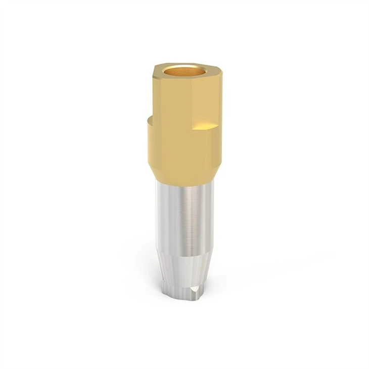
Material | Peek + Titanium alloy |
system | Compatible with Camlog/Ankylos/Teeth/Mis/Dio/Nobel/Osstem/ITI over 30 different systems |
Function | Restoration |
The 3D scan bodies are scanned by scanner to COMPLETE IMPLANT RESTORATION IN EDENTULOUS JAWS
With the 3D dental scanner, The scan bodies apply the Exocad software and under the guidance of the technician, a dental laboratory in Shenzhen successfully achieved the manufacture of full edentulous jaw implant prosthesis through the process of digital dental restoration.
1.Dental scan mode
During the preparation stage, the dental technician in the dental laboratory used 6 scanbodies, and fixed them on analogs inside the stone model with a screwdriver. The artificial gingiva of the model was removed to check whether the connecting pieces were in place or not.
When the scanning step with scanbodies was performed, the artificial gingiva was removed to ensure that the scanbodies were fully seated. After completion of the preparatory work, the upper and lower occlusion models, the mandible model, the artificial gingiva working model and the scanbody working model were successively scanned using the 3D intra-oral scanner under the guidance of your self-contained 3D scanning software DentalScan. When the upper and lower occlusion models were scanned, a special occlusal attachment equipped for the scanner was used. The points marked on the occlusal fixture could be used to identify the spatial position of the model, thus transferring the positional relationship to the CAD design software.
2.Software modeling
Once the 3D scan data was obtained, the technician will design the prosthesis with Exocad software. Within the design software, the technician selected the scanbodies from the CAD data corresponding to the platform size of the implant data and aligned them with the scanbodies section of the scan data successively to achieve the best match. This alignment step is extremely critical, because it would directly determine if the upper structure could achieve a “passive fit”.
The Exocad's best fit function can get the best result by using the CAD data of the scanned bodies in the database to align with the scanned data. As the special bite relationship transfer tool of the 3D scanner was used, the spatial relationship between the maxillary and mandibular models and the dental articulator was completely transferred to the virtual articulator within the design software. Since the special occlusal accessory of the scanner was used, the CNC milling machine virtual articulator could be opened directly within the exocad design software, and the position of the digital model within the virtual articulator was completely consistent with the position of the stone model within the virtual articulator. Therefore, occlusal movement could be fully simulated within the software to eliminate intersections that might appear on the denture during movement.
The occlusal movement is simulated by the virtual articulator in the software. Intersections in the denture can be directly visualized and removed to avoid premature contact in the finished denture.
Based on the full anatomical shape from the previous step, the model is clipped to the basal structure. The mesial-distal retention grooves are prepared with a carving tool. In the processes, the basal bridge was designed using the locations of the mesial-distal retention grooves.
3.Fabrication of the prosthesis
Once the design of the basal bridge was completed, the metal bridge was milled with a five-axis milling machine. The bridge fitted the model very well thanks to many factors, essential among which, however, was the high-quality data acquired by the high-pixel camera.
After processing, the finishing coating with light-curing resin was completed and the aesthetic repair of the red-and-white areas was carried out to obtain very good results.
The processed metal bracket fully fits the model without tilting when trying on. The finish has been made with light-curing resin.
scan bodies for implant is compatible with system below:

scan bodies for implant Pictures


Dental CAD/CAM System Workflow:

Certification:

Packing & Delivery

Company Profile


Exhibition Photo

FAQ
Q: Where is your factory located? How can I visit there?
A: Our factory located in Shenzhen, which is the Hi-Tech developed costal city in southern of China, nearby HongKong and Guangzhou. Half of an hour to HK or Guangzhou by High Railway, or flight to Baoan International Airport. Welcome to our factory for visiting.
Q: How long for the production time ?
A: Normally production time of products is from 3 days to 15 days, depending on the quantity ordered. If you need a rush order, contact our representatives to discuss your specific needs.
Q: What's the delivery time?
A: 5 -7working days by express.
Q: What's the payment method?
A: T/T (Bank Transfer), Western Union, Cash, Paypal.
Q: How can I buy from Chirimen Technology Co.,Ltd?
A: You can send your inquiry to us about the products. Our sales representative will contact you within 24 hours to assist you regarding your inquiry.
Q: What kind of packaging do you offer?
A: Throughout the packing process, preventive measures will be taken so that the goods are in an excellent condition while in transit.
Hot Tags: scan bodies for implant, China, suppliers, manufacturers, factory, wholesale, buy, price, for sale, made in China
Previous
Scanbody ImplantSend Inquiry


